

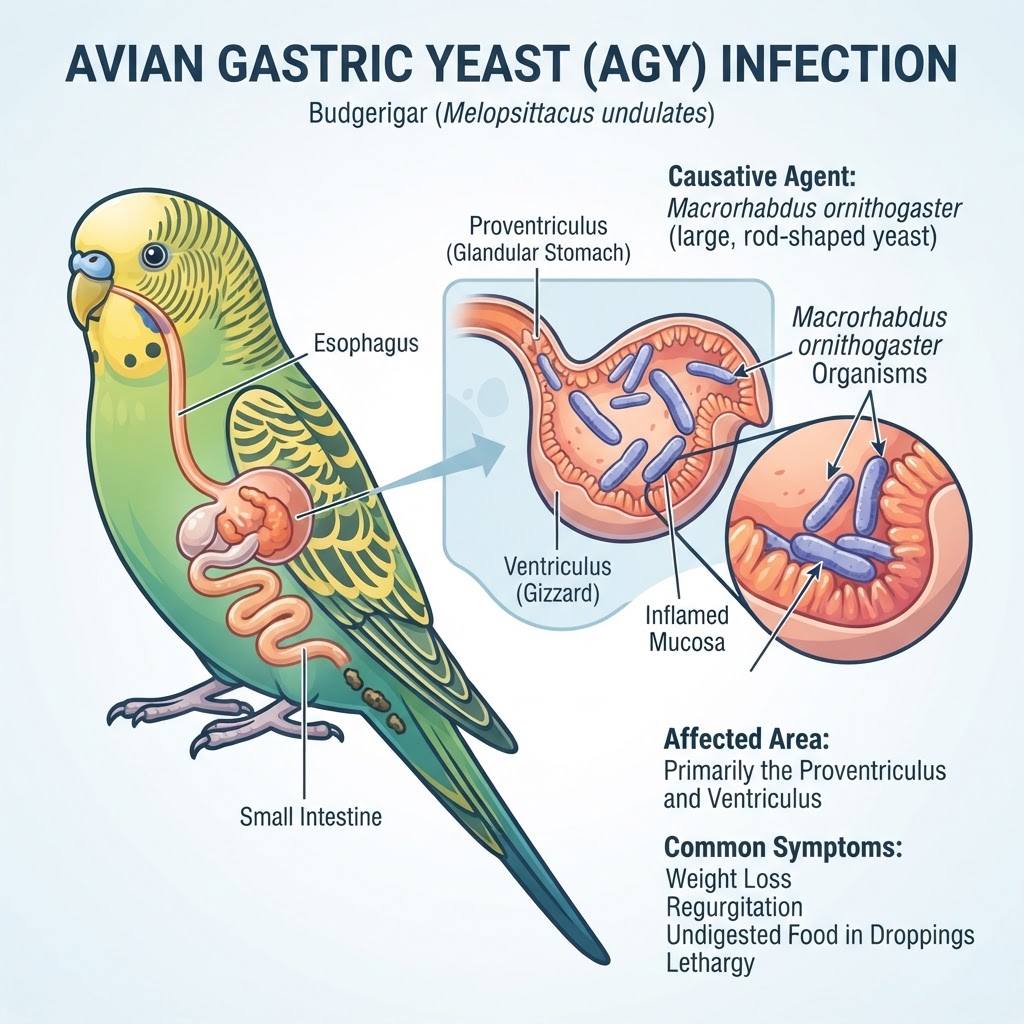
Understanding Macrorhabdus ornithogaster: The Pathophysiology of Avian Gastric Yeast
Avian Gastric Yeast (AGY) is primarily a localized infection of the proventriculus and ventriculus. Unlike typical yeast infections, *Macrorhabdus ornithogaster
- is a large, rod-shaped organism that embeds itself deep within the koilin layer of the stomach lining. This colonization triggers a significant inflammatory response, resulting in the thinning of the stomach wall, glandular atrophy, and in severe cases, deep ulceration.
The Role of pH in Fungal Proliferation
The organism is highly dependent on the acidic environment of the avian stomach. Research suggests that high carbohydrate diets may create an environment that promotes the rapid replication of these organisms. When the mucosal barrier is compromised, the bird loses the ability to properly digest ingesta, leading to the passage of undigested seeds and fibers in the droppings.
Clinical Manifestations and Recognizing Warning Signs
Identifying AGY in the early stages requires keen observation of a bird's daily habits and dropping quality. Because birds are prey animals, they are experts at masking illness until it is advanced.
Early-Stage Indicators
-
Weight loss despite a good appetite: This is a classic sign of the body failing to extract nutrients from food.
-
Regurgitation: Birds may flick their heads or show signs of discomfort after eating.
-
Altered Droppings: Presence of undigested food particles or mucoid urates.
Critical Warning Signs (Emergency Indicators)
If a bird becomes lethargic or displays a fluffed posture, it is in critical condition and requires immediate veterinary intervention. Gastrointestinal stasis caused by the obstruction or severe pain of a ventriculus infection can quickly lead to shock.
Causative Factors and Predisposition
Transmission of AGY is primarily horizontal, occurring through the ingestion of contaminated feces or contaminated food and water sources. The organism is highly resilient and can persist in an aviary environment for extended periods.
Vulnerable Species and Risk Factors
-
Budgerigars: Historically the most commonly affected species.
-
Lovebirds and Cockatiels: Frequently documented as secondary hosts.
-
Stress-Induced Flares: Factors such as exposure to avian viral infections, hormonal shifts, and nutritional deficiencies drastically increase the risk of a subclinical infection becoming symptomatic.
Veterinary Diagnostic Protocols
Veterinary diagnosis relies on a multi-modal approach. Since the organism is not shed continuously, a single negative test result does not rule out infection.
Diagnostic Techniques
-
Fecal Cytology: Using specialized stains to visualize the large, rod-shaped fungi under a microscope.
-
Crop and Proventricular Wash: A sterile saline wash used to recover organisms directly from the site of infection.
-
Imaging (Radiographs): In advanced cases, veterinarians may look for an enlarged proventriculus, which suggests chronic distension.
Therapeutic Interventions and Supportive Care
The primary treatment for AGY is a prolonged course of Amphotericin B. This medication is typically administered orally or mixed into the water supply for a duration of 30 days or more. Because this drug is poorly absorbed in the gastrointestinal tract, it effectively remains in the stomach to target the site of infection.
Managing Secondary Complications
During treatment, birds often require supportive fluid therapy and easily digestible diets to stabilize blood glucose levels. If secondary bacterial enteritis is suspected, broad-spectrum antibiotics may be utilized in conjunction with antifungal therapy.
Recovery, Aftercare, and Long-Term Management
Recovery from AGY is a marathon, not a sprint. The gastrointestinal tract requires time to heal from the erosion caused by the fungus. Post-treatment, many birds continue to require specialized dietary support, including the elimination of excessive sugars and a focus on high-quality, pelleted nutrition to maintain a stable, non-acidic gut environment.
