

Understanding Bovine Respiratory Syncytial Virus Pathogenesis
The Role of Viral Syncytia
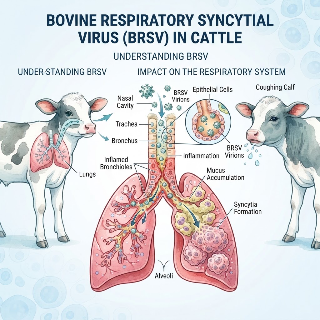
BRSV is an enveloped, single-stranded RNA virus that primarily infects the mucosal surfaces of the upper and lower respiratory tracts. The virus gains entry by attaching to alveolar macrophages and epithelial cells. Once inside, the F-protein on the viral envelope causes neighboring cells to fuse together, creating 'syncytia'—multinucleated giant cells that disrupt normal ciliary clearance and allow Canine Influenza-like spread among herd mates.
Clinical Manifestations and Respiratory Compromise
Recognizing Early Respiratory Signs
Initial infection often manifests as a dry cough, nasal discharge, and a mild fever. As the viral load increases, cattle exhibit signs of respiratory distress, including open-mouth breathing and an exaggerated abdominal effort to draw air into the lungs. If left untreated, the inflammation can progress to interstitial pneumonia, which may be fatal.
Emergency Indicators in Calves and Feedlot Cattle
Emergency veterinary intervention is required if an animal exhibits cyanotic mucous membranes (bluish tint), extreme lethargy, complete anorexia, or severe dyspnea that prevents the animal from lying down. These are signs of end-stage pulmonary compromise often requiring intensive management.
Environmental Factors and Transmission Dynamics
The Impact of Commingling and Stress
BRSV transmission is primarily airborne. In large-scale operations, the introduction of naive stock into a group of chronically infected carrier animals often results in an 'all-in, all-out' transmission cycle. Similar to the transmission dynamics seen with Canine Distemper, the stress of transport, poor ventilation, and extreme weather fluctuations serve as primary catalysts for an outbreak.
Diagnostic Approaches and Veterinary Evaluation
Clinical Examination and Imaging Techniques
Diagnosis is typically based on clinical presentation and history. Veterinarians may use thoracic auscultation to identify harsh bronchial sounds or rales. In complicated cases, ultrasound imaging can be used to visualize consolidation of the lung tissue. For definitive diagnosis, nasal swabs or bronchial alveolar lavage (BAL) samples are sent for PCR testing to identify the viral genome.
Differentiating Respiratory Pathogens
Because clinical signs often overlap with Canine Parvovirus (in a respiratory-distress context) or bacterial pneumonias, clinicians must distinguish BRSV from infectious bovine rhinotracheitis (IBR) or bovine viral diarrhea (BVD).
Therapeutic Intervention and Supportive Care
Managing Pulmonary Inflammation
Treatment focuses on supportive care and preventing the transition to secondary bacterial pneumonia. Non-steroidal anti-inflammatory drugs (NSAIDs) are commonly administered to reduce pulmonary edema and fever. In high-risk cases, veterinarians may prescribe broad-spectrum antibiotics to address the secondary bacterial infections that frequently follow BRSV damage.
Supportive Hydration and Environmental Control
Providing a clean, dry, and dust-free environment is critical. In cases of dehydration or anorexia, intravenous fluid therapy may be necessary to maintain metabolic function.
Herd Immunity and Prevention Strategies
Vaccine Efficacy and Vaccination Timing
Vaccination is the cornerstone of prevention. Both modified-live and killed vaccines are available. Strategic vaccination must be performed well in advance of high-stress events, such as weaning or transport, to ensure the animal has developed a robust neutralizing antibody titer.
