

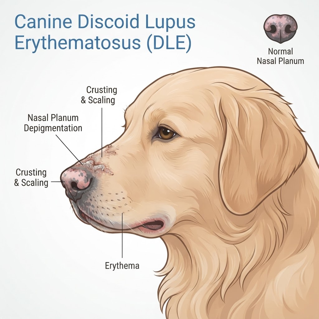
Understanding Discoid Lupus Erythematosus in Canines
Discoid lupus erythematosus (DLE) is a localized form of autoimmune disease in dogs that primarily manifests as chronic inflammation of the skin. It is characterized by the body’s inability to distinguish its own keratinocytes from foreign invaders, leading to a localized immune assault at the dermal-epidermal junction. While it is rarely life-threatening, it is a significant source of chronic discomfort for affected pets and may be confused with Nasal Planum Hyperkeratosis by owners unfamiliar with autoimmune skin conditions.
Clinical Presentation and Progression
The initial changes associated with DLE are often cosmetic and subtle. The dark pigment of the nose may fade to a pale pink or gray color, and the characteristic pebbled texture of the skin may smooth out. As the condition advances, the skin becomes fragile, developing scales, crusts, and shallow ulcers. In some cases, the tissue loss can lead to permanent scarring or architectural deformity of the nose.
Identifying Signs of Nasal and Facial Dermatitis
Recognizing DLE involves monitoring for persistent changes in skin appearance around the face. Unlike Atopic Dermatitis, which typically presents with generalized pruritus and paw licking, DLE is site-specific.
Warning Signs of Disease Escalation
If the lesions begin to ooze, bleed, or exhibit a foul odor, this often indicates secondary bacterial infection. While many skin conditions might look similar, persistent crusting that fails to resolve with simple moisturizing or barrier ointments is a clinical red flag. Owners should also watch for increased sensitivity to touch, as ulcerated skin can become extremely painful and prone to secondary inflammation akin to Fly Bite Dermatitis in terms of localized irritation.
The Role of Ultraviolet Light and Genetic Predisposition
While DLE is an autoimmune condition, it is strongly photo-aggravated. Ultraviolet radiation alters the antigenicity of the skin, triggering the immune system to intensify its attack.
Breed and Environmental Factors
Certain breeds appear more susceptible, including Collies, Shetland Sheepdogs, and German Shepherds. Unlike Zinc-Responsive Dermatosis, which is linked to nutritional uptake, DLE has a clear genetic correlation with immune system regulation. Managing exposure to direct sunlight is the most effective environmental modification strategy to limit the exacerbation of symptoms.
Veterinary Diagnostic Protocols
Veterinarians must differentiate DLE from other ulcerative skin conditions through specific testing. This usually starts with a physical examination to rule out systemic illnesses or metabolic issues like Vitamin A-Responsive Dermatosis.
Histopathology and Skin Biopsies
The gold standard for diagnosing DLE is a skin biopsy. A pathologist will look for interface dermatitis, where lymphocytes infiltrate the basement membrane, and signs of vacuolar degeneration of the basal cells. In some instances, specialized immunofluorescence staining may be used to confirm the presence of immune complexes.
Therapeutic Management Strategies
The goal of treatment is to suppress the local immune response and protect the skin from further damage. Medications typically include topical steroids or immune-modulating creams such as tacrolimus. In cases where the condition is more aggressive, systemic medications like doxycycline, niacinamide, or oral corticosteroids may be required.
Long-Term Maintenance and UV Protection
Lifelong management is required to keep the condition in remission. Regular applications of high-SPF, dog-safe sunscreen are essential during outdoor activities. If the condition is severe or unresponsive, veterinarians may discuss alternative therapies such as cyclosporine or hydroxychloroquine to maintain skin integrity and prevent the need for more invasive dermatological interventions.
