

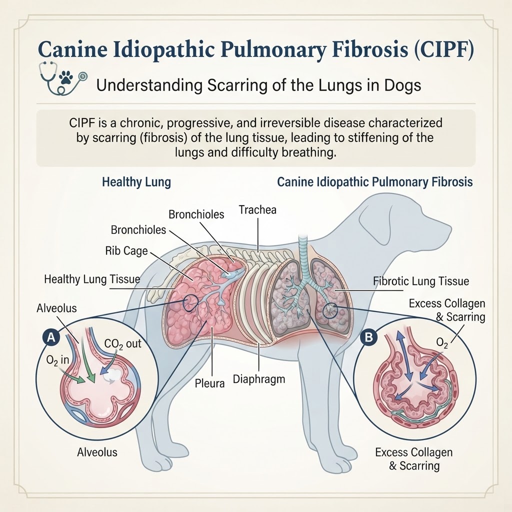
Understanding Pulmonary Fibrosis and Alveolar Interstitial Disease
Canine idiopathic pulmonary fibrosis represents a complex interstitial lung disease where the normal lung architecture is replaced by dense, non-functional fibrous tissue. In a healthy canine lung, the alveolar-capillary membrane is thin and efficient, allowing for rapid gas exchange. In CIPF, this membrane thickens significantly due to the deposition of collagen and other extracellular matrix components. This process essentially turns the soft, spongy lung tissue into a stiff, inelastic structure that resists expansion during inhalation.
The Role of Pulmonary Interstitium Damage
The disease process involves repeated cycles of injury to the epithelial cells lining the air sacs. Although the exact trigger for this injury remains unknown, the body's attempt to repair these micro-injuries leads to the chronic, maladaptive deposition of fibrous scar tissue. This is fundamentally different from a simple respiratory infection or acute asthma; it is a permanent architectural change that cannot be reversed by antibiotics or standard anti-inflammatory medications.
Recognizing Clinical Indicators and Respiratory Distress
The primary hallmark of CIPF is the onset of chronic, non-productive coughing and progressive exercise intolerance. Owners often notice that a once-active dog begins to tire prematurely during walks or struggles to keep up with household activity.
- Exertional Dyspnea: Difficulty breathing during even light exercise.
- Inspiratory Crackles: A signature 'velcro-like' sound heard during a vet's auscultation, caused by the snapping open of collapsed, fibrotic airways.
- Tachypnea: An increased resting respiratory rate as the body struggles to maintain oxygen levels.
- Cyanotic Mucous Membranes: A bluish tint to the gums indicating severe hypoxemia (low oxygen in the blood).
- Syncope: Brief fainting spells, often secondary to the heart struggling to pump through hardened, restricted lung tissue.
Emergency Warning Signs
If you observe your dog experiencing extreme difficulty breathing, such as open-mouth breathing at rest, extreme lethargy, or if the tongue turns blue, this constitutes a medical emergency. Unlike diabetes mellitus, which can be managed with daily insulin, or hypothyroidism, which responds to hormone replacement, respiratory failure in CIPF can reach a critical point very rapidly, necessitating immediate supplemental oxygen therapy.
Etiology and Breed-Specific Risk Factors
While the condition is labeled 'idiopathic,' genetic studies strongly point to a hereditary predisposition in certain terrier breeds. The condition is most famously associated with the West Highland White Terrier, but it has been documented in other breeds like the Cairn Terrier, Staffordshire Bull Terrier, and occasionally in older, larger-breed dogs. The onset usually occurs in middle-aged to senior dogs, rarely appearing before the age of 8 or 9.
Genetic vs. Environmental Contributions
Researchers are investigating whether chronic exposure to inhaled pollutants or low-level, long-term inflammation contributes to the progression in genetically susceptible individuals. It is not currently linked to autoimmune conditions like Addison's disease, though systemic health is paramount in supporting a dog with compromised lung function. Unlike chronic kidney disease, which can be partially managed through strict dietary phosphorus control, there is no known 'fibrosis-friendly' diet, though maintaining an optimal weight is vital to reduce the work of breathing.
Diagnostic Pathways in Veterinary Cardiology and Pulmonology
Diagnosing CIPF is a process of exclusion. Because the signs overlap with heart disease or chronic bronchitis, your veterinarian will typically begin with a thorough physical examination followed by thoracic radiography. Radiographs of a dog with CIPF often show a 'bronchointerstitial pattern,' which can sometimes be mistaken for Cushing's syndrome systemic effects on the lungs or primary cardiac disease.
Advanced Imaging and Lung Assessment
- Thoracic Radiography: Used to rule out heart failure or masses.
- Echocardiography: Crucial to ensure the coughing is not being caused by mitral valve disease or secondary pulmonary hypertension.
- CT Scan (High-Resolution): The gold standard for visualizing the 'ground glass' appearance of fibrotic lung tissue.
- Bronchoalveolar Lavage (BAL): Used to collect fluid samples from the deep lung to rule out inflammatory bowel disease secondary complications or primary infections.
Multimodal Management and Palliative Care
Because no cure exists, treatment focuses on optimizing comfort and oxygen utilization. Bronchodilators, such as theophylline or terbutaline, are often prescribed to widen the airways and facilitate easier airflow. In cases where significant inflammation is present, low-dose corticosteroids may be used, though their efficacy is debated.
Managing Secondary Complications
If secondary pulmonary hypertension is confirmed via echocardiogram, medications like sildenafil (Viagra) are frequently utilized to reduce the pressure in the blood vessels surrounding the lungs, which helps improve the dog's endurance. Supportive care also includes limiting strenuous exercise and ensuring a low-stress, climate-controlled environment to prevent panting, which puts further stress on the respiratory system.
Long-Term Outlook and Supportive Care Strategies
Living with a dog with CIPF requires vigilance. You must monitor their respiratory rate while sleeping; a resting rate consistently over 30 breaths per minute is often a trigger to contact your veterinarian. Keeping the environment free of dust, smoke, and strong aerosol sprays is essential, as these can irritate the already compromised airways.
Unlike managing cognitive dysfunction syndrome, which involves lifestyle adjustments for mental clarity, managing CIPF is about physical limitations. Owners should work closely with a veterinary internist to adjust medications as the disease progresses and ensure that comfort measures, such as portable oxygen concentrators, are considered if the disease reaches a severe stage.
