

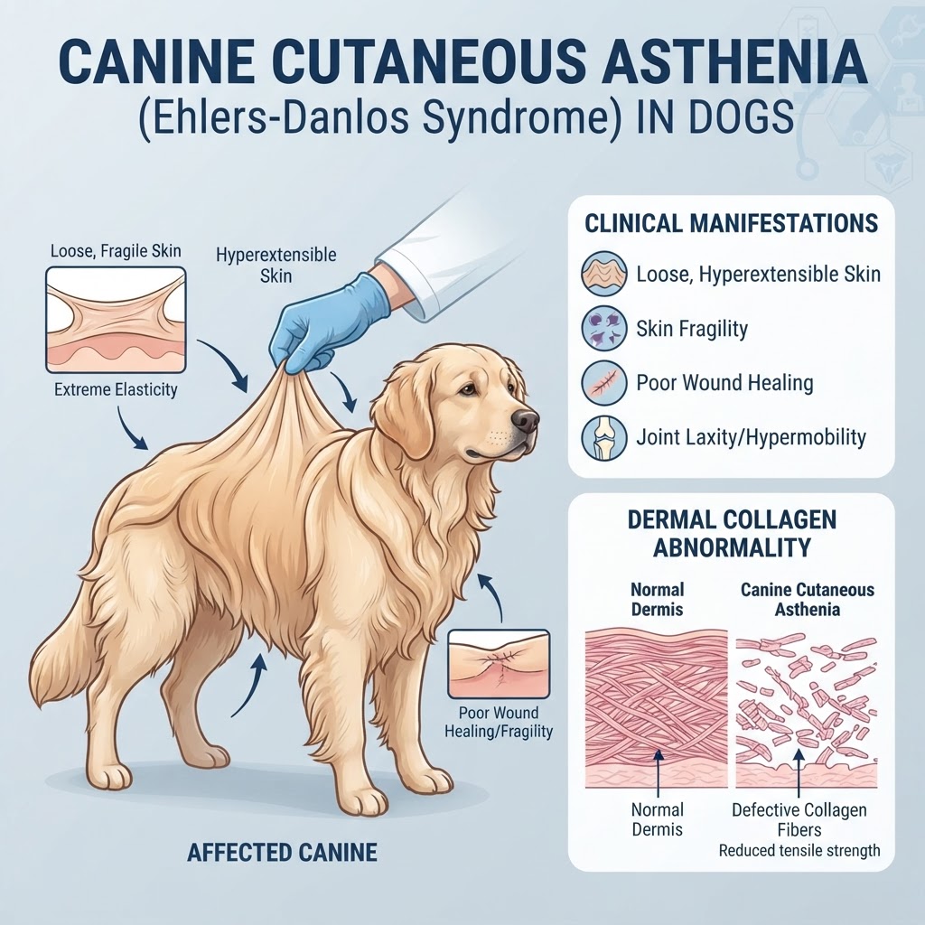
Understanding Ehlers-Danlos Syndrome: The Pathophysiology of Cutaneous Asthenia
Canine cutaneous asthenia, often classified as an Ehlers-Danlos syndrome, is an inherited defect of connective tissue. At the heart of this condition is a fundamental failure in the production of healthy, strong collagen fibers. Collagen serves as the 'glue' that holds the dermis, tendons, and ligaments together. In affected dogs, this glue is effectively weakened, leading to a state of extreme fragility in the skin.
Biochemical Mechanisms and Collagen Synthesis
In a healthy canine, collagen proteins interweave to create a durable, flexible matrix. Dogs with this condition often lack the necessary enzymes or structural blueprints to create this matrix correctly. This results in thin, hyper-extensible skin. Because the vascular support within the skin is also built upon this collagen, the blood vessels are often fragile, leading to significant bleeding or bruising upon minor impact. This condition is frequently compared to Von Willebrand Disease in terms of the clinical concern for unexplained bleeding, though the etiology of the vascular fragility in cutaneous asthenia is structural rather than coagulation-based.
Identifying Clinical Signs and Warning Signs of Fragility
Recognizing the condition often happens when a pet parent notices that the skin seems 'too big' or feels like velvet to the touch. The skin lacks the usual firmness and structural attachment to the underlying muscle and fat layers.
Physical Manifestations of Dermatosparaxis
-
Skin Hyper-extensibility: The skin can be pulled significantly away from the body before reaching its limit.
-
Spontaneous Lacerations: Tears may occur without any clear traumatic event, particularly over the back, neck, or tail.
-
Hematoma Formation: Easy bruising or pockets of blood occurring after very mild pressure.
-
Joint Hypermobility: Excessive range of motion in joints, which can lead to early-onset Canine Hip Dysplasia or instability.
-
Delayed Wound Healing: Surgical sites and accidental wounds often dehisce (tear open) because the skin cannot hold sutures.
When to Seek Emergency Care
If you notice large, gaping wounds that do not stop bleeding, or if your dog displays signs of sudden lameness or joint pain that may be related to Patellar Luxation due to ligamentous laxity, emergency intervention is required. Constant skin monitoring is essential to prevent secondary infections.
Etiology: The Genetic Basis of Connective Tissue Defects
Cutaneous asthenia is largely genetic, typically inherited in an autosomal recessive or autosomal dominant pattern, depending on the specific breed. The condition stems from mutations in genes responsible for the production of types I, III, or V collagen.
Breed-Specific Predispositions
While any dog can be affected, certain breeds show a higher prevalence. Breeds often associated with collagen-related abnormalities include the Greyhound, Boxer, Springer Spaniel, and German Shepherd. In these breeds, the trait may be linked to specific bloodlines, making selective breeding essential to reducing the occurrence of the disorder. Like Elbow Dysplasia, this is a structural, developmental disease that requires careful management throughout the dog’s life.
Diagnostic Approaches and Veterinary Evaluation
Diagnosing cutaneous asthenia requires a combination of physical examination and, occasionally, specialized skin biopsy.
The Skin Extensibility Index (SEI)
A veterinarian will perform a physical exam to measure the extensibility of the skin over the dorsum. This index compares the skin pull to the animal's length. Imaging, such as radiographs, is often utilized to rule out co-occurring orthopedic issues or to evaluate the degree of joint laxity. Laboratory work, including coagulation panels, is recommended to differentiate this condition from bleeding disorders, ensuring that a simple nick isn't masking a deeper clotting issue like Hemophilia A.
Managing Chronic Fragility: Treatment and Long-Term Care
There is no known cure for cutaneous asthenia, as it is a permanent structural defect of the body’s cells. Treatment is focused on environmental modification, trauma prevention, and supportive care.
Surgical Considerations and Wound Management
Surgical intervention is highly discouraged unless strictly necessary due to the extreme risk of skin failure post-operatively. When surgery is required, specialized suturing techniques—such as tension-relieving mattress sutures—must be employed. Long-term management involves the daily use of protective clothing to prevent environmental trauma and keeping the dog away from rough surfaces, dense brush, or other dogs that play too aggressively.
