

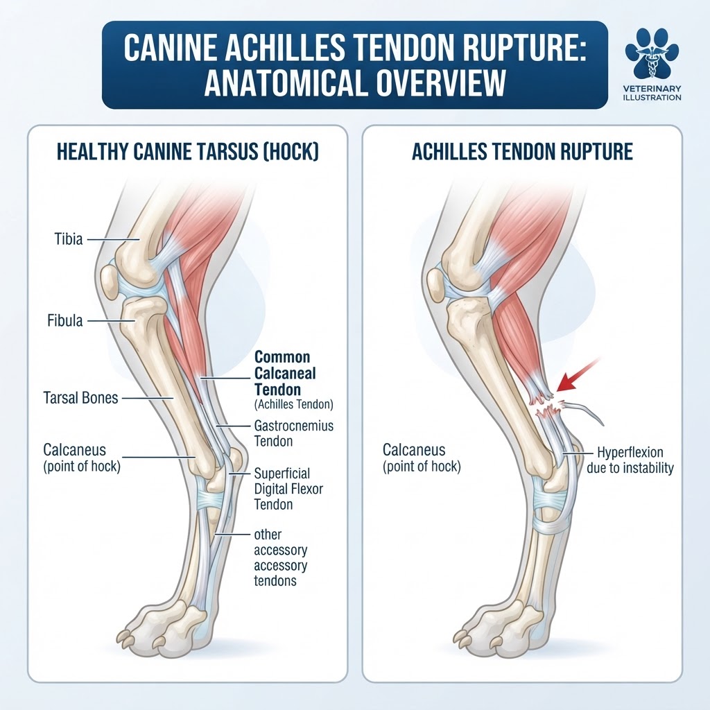
Understanding Achilles Tendon Rupture in Canines
An Achilles tendon rupture, more accurately termed a calcaneal tendon rupture, is a severe injury affecting the primary extensor mechanism of the canine hindlimb. This traumatic event involves the disruption of the calcaneal tendon, a robust band of fibrous connective tissue that anchors the powerful calf muscles to the heel bone (calcaneus). This tendon is crucial for normal locomotion, enabling dogs to walk, run, and jump by extending the hock joint. When rupture occurs, this biomechanical function is compromised, leading to immediate and often profound lameness.
Types of Achilles Tendon Rupture
Achilles tendon ruptures in dogs are typically categorized by the extent of the damage:
- Complete Rupture: The tendon is fully severed, resulting in a total loss of its functional integrity. This is the most common and severe form.
- Partial Rupture: The tendon fibers are torn but not completely separated. While less severe than a complete rupture, it still leads to significant lameness and instability.
Recognizing Signs of Achilles Tendon Rupture
The onset of symptoms is usually sudden and dramatic, often following a specific incident like a fall, jump, or impact. Owners should be vigilant for the following signs:
- Acute, Severe Lameness: The most prominent sign is an immediate and significant limp in one or both hindlimbs, with the dog refusing to bear weight on the affected leg(s).
- Abnormal Hock Angle (Dropped Hock): The hock joint appears to 'collapse' inward and backward, losing its normal rigid extension. The dog may be observed walking on the back of its hock or fetlock.
- Inability to Stand or Walk Normally: The dog may struggle to rise, remain in a sitting or lying position for extended periods, or exhibit a very unstable, wobbly gait when it does move.
- Swelling Around the Hock: Inflammation can cause noticeable swelling on the back of the hock joint where the tendon is located.
- Palpable Gap: In cases of complete rupture, a veterinarian may be able to feel a gap or discontinuity in the tendon's structure.
- Audible Pop or Snap: Some dogs may vocalize or exhibit a sound at the moment of injury, indicating a sudden, forceful tear.
Emergency Warning Signs: While lameness is always concerning, a complete inability to bear weight on a hindlimb and a visibly 'dropped' or unstable hock joint are immediate indicators that veterinary emergency care is required.
Etiology and Predisposing Factors
The causes of Achilles tendon rupture are primarily traumatic, though underlying degenerative changes can increase susceptibility.
Traumatic Causes
Direct trauma is the most common culprit. This includes:
- High-Impact Activity: Sudden stops, jumps from heights (e.g., off furniture or into a vehicle), or twisting motions during play can overstretch and tear the tendon.
- Direct Blows: Accidents involving impacts to the back of the hindlimb, such as being hit by a car, gunshot injury, or severe bite wounds.
- Falls: Slipping on slick surfaces or during strenuous activity can place excessive strain on the tendon.
Degenerative Changes and Risk Factors
While trauma is often the initiating event, the tendon's condition at the time of injury plays a significant role:
- Tendinopathy: Chronic inflammation or degeneration of the tendon (tendinopathy) weakens its structure, making it more prone to rupture even with less severe trauma. This can be age-related or due to repetitive stress.
- Steroid Use: Long-term or high-dose corticosteroid administration has been linked to weakening of connective tissues, including tendons, potentially increasing rupture risk.
- Certain Medications: Some medications might have adverse effects on tendon health, although this is less common.
Breed Predisposition in Canine Achilles Tendon Rupture
While Achilles tendon rupture can affect any dog, certain breeds may be more predisposed due to their conformation, activity levels, or genetic factors. Some studies suggest a higher incidence in large breeds, but it is not as strongly breed-specific as some other orthopedic injuries like cruciate ligament rupture or hip dysplasia. However, breeds known for high agility and jumping activities might experience it more frequently due to the nature of their use.
Diagnostic Approach in Veterinary Medicine
Diagnosing an Achilles tendon rupture involves a thorough physical examination and often imaging studies to confirm the injury and assess its severity.
The Physical Examination
During a physical exam, the veterinarian will meticulously assess the affected limb. Key findings include:
- Gait Analysis: Observing the dog walk and stand to document the lameness and abnormal posture.
- Palpation: Gently feeling the back of the hock to assess for swelling, heat, pain, and any discontinuity or gap in the calcaneal tendon.
- Passive Range of Motion: Evaluating the movement of the hock and stifle joints. With a ruptured Achilles tendon, the hock may appear excessively mobile or unstable in an unnatural direction.
- Thompson Test: This specific test involves squeezing the calf muscles. In a healthy dog, this action causes plantarflexion (extension) of the hock. In a dog with a ruptured Achilles tendon, there will be little to no hock movement because the tendon is not intact to transmit the muscle contraction.
Diagnostic Imaging
- Radiographs (X-rays): While X-rays do not directly visualize soft tissues like tendons, they are crucial for ruling out fractures or dislocations of the hock or other bones that could cause similar lameness. They can also indirectly suggest tendon issues if there is displacement of the calcaneus bone.
- Ultrasound: This is the gold standard for diagnosing tendon injuries. A high-frequency ultrasound can directly visualize the tendon fibers, showing the site of rupture, the extent of tearing (partial vs. complete), and any associated inflammation or fluid accumulation.
- MRI/CT Scan: In complex cases or when ultrasound is inconclusive, Magnetic Resonance Imaging (MRI) or Computed Tomography (CT) may be used, though these are less common for routine Achilles tendon rupture diagnosis due to cost and availability.
Laboratory Work
Blood work (complete blood count and chemistry panel) is typically performed prior to anesthesia for surgical repair. This helps assess the dog's overall health, organ function, and suitability for surgery and anesthesia. It does not directly aid in diagnosing the tendon rupture itself but is essential for pre-operative screening.
Treatment Strategies for Achilles Tendon Rupture
Treatment for Achilles tendon rupture typically involves surgical repair to restore the tendon's integrity and function. Conservative management is rarely successful for complete ruptures.
Surgical Intervention
The primary goal of surgery is to reattach the ruptured tendon ends or to reconstruct a functional tendon using surrounding tissues or implants.
- Tendon Suture/Repair: The torn ends of the calcaneal tendon are directly suton to be reapproximated and sutured together using strong, specialized suture materials. This is often performed in cases of complete rupture where sufficient tendon length remains.
- Tendon Transposition/Reconstruction: In cases where tendon ends are damaged or missing, the surgeon may use adjacent tendons (like the superficial digital flexor tendon) or create a new 'tendon' from available tissue to bridge the gap and reattach to the calcaneus.
- Internal Fixation: Sometimes, implants like screws or pins might be used to stabilize the calcaneus bone during the healing process, especially if there are associated bone fragments.
Medications and Supportive Care
- Pain Management: Post-operatively, pain medication (NSAIDs, opioids) is crucial to manage discomfort and inflammation.
- Antibiotics: Administered peri-operatively to prevent wound infection.
- Activity Restriction: Strict rest is paramount during the initial healing phase. This involves confinement to a crate or small room, with leash-controlled potty breaks only.
- Splinting/Casting: In some cases, a protective splint or cast may be applied to immobilize the limb and protect the surgical repair, especially during the early stages of healing.
Recovery and Post-Operative Management
Recovery from Achilles tendon rupture repair is a lengthy process requiring significant owner commitment. The timeline is divided into distinct phases:
Phase 1: Initial Healing and Immobilization
- Timeframe: 0-4 weeks post-surgery.
- Description: This critical phase focuses on protecting the surgical repair. Activity is strictly limited to short, leashed potty breaks. Sutures are typically removed around 10-14 days. A splint or cast may be used during this period to maintain immobility and protect the repair site. Pain management is essential.
Phase 2: Gradual Weight-Bearing and Controlled Movement
- Timeframe: 4-8 weeks post-surgery.
- Description: If the initial healing is progressing well (as confirmed by veterinary recheck), activity restrictions are gradually eased. Leash walks are slowly increased in duration and frequency. Passive range of motion exercises may be introduced. The splint/cast is typically removed if used.
Phase 3: Strengthening and Return to Activity
- Timeframe: 8-16 weeks (and beyond) post-surgery.
- Description: Dogs begin to bear more weight on the leg. Controlled exercises, such as walking on uneven surfaces, walking up/down gentle inclines, and short periods of off-leash movement in a controlled area, are introduced. The goal is to rebuild muscle mass and strength in the affected limb.
Phase 4: Full Recovery and Return to Normal Function
- Timeframe: 4-6 months or longer.
- Description: Most dogs can gradually return to normal activities, including running and playing, provided there are no signs of pain or instability. Long-term monitoring for any signs of re-injury or chronic changes is important. Full function may take up to a year to achieve in some cases.
Prevention Strategies for Achilles Tendon Injuries
While not all Achilles tendon ruptures can be prevented, especially those resulting from severe, unavoidable trauma, certain measures can reduce the risk:
- Maintain Healthy Weight: Obesity places undue stress on joints and tendons.
- Appropriate Exercise: Ensure dogs receive regular, consistent exercise suitable for their age, breed, and fitness level. Avoid sudden bursts of intense activity if the dog is not conditioned.
- Safe Environment: Minimize risks by ensuring safe play areas, avoiding slippery surfaces, and being cautious with jumps from heights.
- Prompt Care for Injuries: Address any minor limping or leg injuries immediately to prevent them from worsening or leading to compensatory problems.
- Awareness of Medications: Discuss any concerns about medications that might affect connective tissue health with your veterinarian.
When to Seek Emergency Veterinary Care
An Achilles tendon rupture is a surgical emergency. Owners should seek immediate veterinary attention if their dog exhibits any of the following:
- Sudden, Severe Lameness: Especially if the dog cannot bear any weight on a hindlimb.
- Visible Abnormal Hock Position: A 'dropped hock' or visibly unstable, collapsing joint.
- Suspected Trauma: If the lameness occurs after a known traumatic event (fall, impact, fight).
- Signs of Shock: Pale gums, rapid breathing, weakness, or collapse, which could indicate a more severe systemic injury accompanying the tendon rupture.
Do not delay seeking care, as prompt diagnosis and surgical intervention significantly improve the chances of a successful recovery and return to function.
