

Understanding Ocular Proptosis and Orbital Trauma
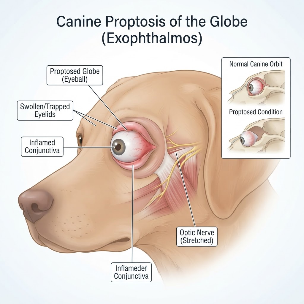
Proptosis of the globe, specifically known as traumatic ocular proptosis, is a condition where the eyeball is forced out of the bony orbit. This occurs due to external trauma or pressure applied to the head and neck. In dogs, this condition is particularly common in brachycephalic (short-nosed) breeds, such as Pugs, Shih Tzus, and Pekingese, because their shallow eye sockets and prominent globes offer less natural protection against blunt force.
Brachycephalic Ocular Syndrome and Anatomical Predisposition
These breeds often suffer from what is known as brachycephalic ocular syndrome, which includes eyelid abnormalities and shallow orbital depths. When a dog experiences a sudden impact—such as a fall, a bite wounds event, or even significant pulling on a leash near the head—the orbital contents are pushed forward. Because the eyelids are structurally incapable of closing behind the globe once it passes the orbital rim, they become stuck, preventing the eye from retracting.
Recognizing Clinical Signs and Emergency Indicators
Recognizing the signs of a proptosed globe is straightforward, but the urgency cannot be overstated. Owners will observe a bulging eye that is fixed in a protruding position. Because the eye cannot blink, it becomes dry and inflamed within minutes.
Ocular and Systemic Warning Signs
- Globe Displacement: The eye is clearly visible and protruding from the eyelid margins.
- Eyelid Entrapment: The eyelids are flipped inside out or stuck behind the globe.
- Corneal Desiccation: The surface of the eye appears dull, dry, or crusty due to exposure.
- Conjunctival Chemosis: Intense, balloon-like swelling of the pink tissue around the eye.
- Hyphema: Blood collecting in the anterior chamber of the eye due to trauma.
- Mydriasis: A fixed or dilated pupil, which is a major indicator of optic nerve damage.
Etiology: Causes and Risk Factors
Proptosis is almost exclusively the result of blunt force trauma. While the breeds mentioned above are predisposed, any dog can experience this if the force is sufficient. In some cases, a crush injury to the head can cause secondary proptosis. It is critical to differentiate this from retrobulbar masses or abscesses that push the eye forward slowly, though these rarely result in the acute "trapping" seen in sudden traumatic events.
Veterinary Diagnostic Procedures
When a dog arrives at the clinic with a suspected proptosis, the veterinarian acts immediately to stabilize the patient. Diagnosis is based on a physical examination of the ocular anatomy. The veterinarian will assess the pupil size and light response (the pupillary light reflex), which are key prognostic indicators for vision retention. A fluorescein stain may be used to check for severe corneal damage, and ultrasound of the orbit may be conducted to rule out fractures or retrobulbar hemorrhage.
Surgical Repositioning and Medical Management
Treatment follows a strict protocol. Initially, the eye must be kept moist with saline or lubricating ointments. If the eye has been out for a short duration, the veterinarian may attempt a lateral canthotomy to widen the eyelid opening, allowing the globe to be gently massaged back into the orbit. Once replaced, a temporary tarsorrhaphy (suturing the eyelids closed) is performed to keep the globe in place while inflammation subsides.
Post-Surgical Considerations
After surgery, the patient is placed on intensive medical management, including:
- Systemic Antibiotics: To prevent wound infection in the damaged orbital tissues.
- Anti-inflammatories: Corticosteroids are often used to reduce orbital swelling.
- Topical Ocular Medication: Antibiotic drops and lubricants to heal the corneal surface.
Recovery, Prognosis, and Long-Term Care
The recovery timeline depends on the severity of the damage. Most patients remain on strict rest for 2-4 weeks. Owners must be prepared for the possibility of enucleation if the eye becomes phthisical (shrinks and becomes painful) or if the damage to the optic nerve is permanent. Regular monitoring of the intraocular pressure and corneal health is essential in the months following the incident.
