

Understanding Meniscal Tears in Canine Stifle Joints
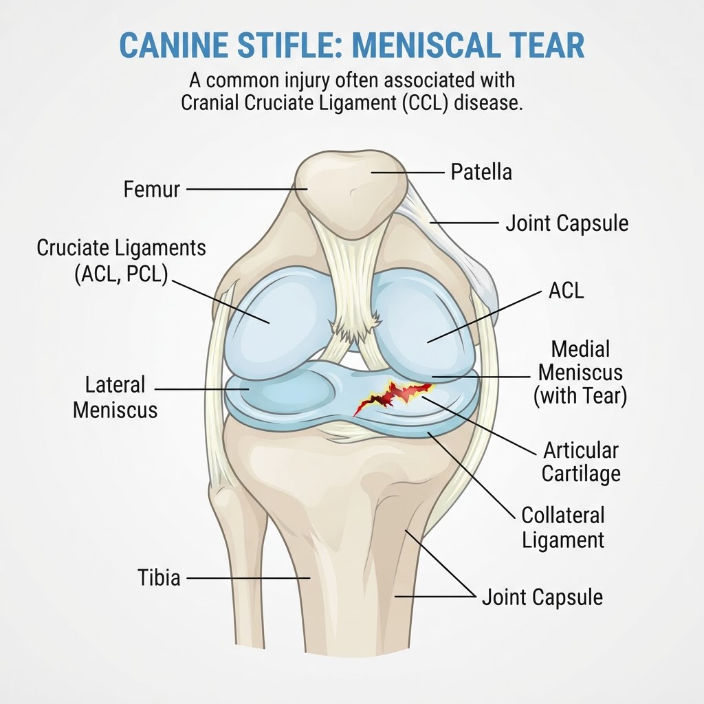
A meniscal tear refers to damage to the cartilage pads, known as menisci, located within the knee (stifle) joint of a dog. These C-shaped structures serve as crucial shock absorbers and stabilizers between the femur and tibia. When injured, they can significantly impact a dog's mobility and comfort.
Types and Pathophysiology of Meniscal Injury
Meniscal tears in dogs are primarily categorized by the mechanism of injury and the appearance of the tear. The two menisci are the medial meniscus, located on the inner side of the knee, and the lateral meniscus, on the outer side. The medial meniscus is more frequently involved in injuries, often due to its structural attachments and role in stabilizing the joint, particularly when the anterior cruciate ligament is compromised.
Pathologically, tears can be classified as acute or degenerative. Acute tears are sudden, often resulting from a specific traumatic event like a fall, jump, or forceful twist. Degenerative tears occur over time due to wear and tear, often in older dogs or those with pre-existing joint conditions such as osteoarthritis. These degenerative changes weaken the meniscus, making it more susceptible to tearing even with minor stress.
Common tear patterns include:
- Longitudinal tears: Running along the length of the meniscus.
- Radial tears: Across the width of the meniscus.
- Bucket-handle tears: A large segment of the meniscus is detached.
- Horizontal cleavage tears: Separating the upper and lower layers of the meniscus.
- Complex tears: Combinations of the above.
These tears can lead to fragments of cartilage becoming loose within the joint, causing irritation, inflammation, and pain. The instability created by the tear can also lead to progressive damage to the articular cartilage, a hallmark of osteoarthritis.
Recognizing Meniscal Tear Symptoms in Dogs
The clinical signs of a meniscal tear can vary depending on the severity and location of the injury, as well as whether it's an acute or chronic issue. Owners often notice a sudden onset of lameness, particularly after strenuous activity or a perceived injury.
Common Signs of Meniscal Dysfunction
- Intermittent or persistent lameness: This is the most common sign. The lameness may improve with rest but often returns with activity. It may be more pronounced on the affected hind limb.
- "Skipping" or "Hitching" Gait: Dogs may take a few steps normally, then "skip" or "hitch" the affected leg, bringing it forward quickly before placing weight on it.
- Stiffness after rest: The dog may appear stiff and reluctant to move immediately after lying down, with mobility improving after a short period.
- Pain on manipulation: The veterinarian may elicit pain when flexing or extending the affected stifle joint.
- "Clicking" or "Popping" Sound: A palpable or audible "click" may be felt or heard during stifle joint manipulation, indicative of displaced meniscal fragments.
- Reluctance to bear weight: In severe cases, the dog may refuse to put any weight on the affected limb.
- Muscle Atrophy: Over time, disuse of the limb can lead to a noticeable decrease in muscle mass in the thigh.
- Swelling: While not always obvious externally, mild swelling within the joint capsule may be present.
Factors Contributing to Meniscal Tears
Meniscal tears are often multifactorial, involving a combination of predisposing factors and acute or chronic stressors.
Predisposing Factors and Risk Identification
- Anterior Cruciate Ligament (ACL) Rupture: This is a very common precursor. A partial or complete rupture of the ACL often leads to abnormal joint motion and increased stress on the menisci, frequently resulting in a secondary meniscal tear, particularly the medial meniscus.
- Degenerative Joint Disease (Osteoarthritis): Chronic inflammation and wear-and-tear in the joint can weaken the meniscal cartilage, making it prone to tearing. Many dogs diagnosed with meniscal tears also have underlying osteoarthritis.
- Trauma: Direct trauma, such as a fall during exercise, a sudden stop and twist, or impact from an accident, can cause acute meniscal tears. This can occur during play, agility training, or even just running in the yard.
- Obesity: Excess body weight significantly increases the load on the stifle joint, accelerating degenerative changes and increasing the risk of meniscal injury.
- Genetics and Breed Predispositions: While not as definitively linked as with some other orthopedic conditions, certain breeds may have a higher propensity for knee issues. However, any breed can experience meniscal tears.
- Age: Older dogs are more susceptible to degenerative tears due to the natural aging process of cartilage.
- Conformation: Abnormalities in limb alignment or joint structure may contribute to uneven stress distribution within the stifle.
Diagnosing Meniscal Tears in Canine Patients
Diagnosing a meniscal tear typically involves a thorough physical examination, diagnostic imaging, and sometimes exploratory surgery.
The Veterinary Diagnostic Approach
-
History and Physical Examination: The veterinarian will begin by discussing the onset of lameness and any observed symptoms. A detailed physical examination of the stifle joint is crucial. This involves palpation of the joint for pain, swelling, and instability. Specific orthopedic tests, such as the cranial tibial thrust and medial buttress test, are performed to assess for signs of ligamentous instability that might suggest an ACL rupture, often associated with meniscal tears. The presence of a "click" during range of motion is a key finding.
-
Radiographs (X-rays): While X-rays cannot directly visualize the menisci, they are essential for evaluating the overall health of the stifle joint. Radiographs can identify signs of osteoarthritis, such as joint effusion (fluid buildup), sclerosis (bone hardening), and osteophytes (bone spurs), which are often present concurrently. They also help rule out other causes of lameness, such as fractures or hip dysplasia.
-
Advanced Imaging: In some cases, advanced imaging techniques may be considered:
-
Magnetic Resonance Imaging (MRI): MRI offers detailed visualization of soft tissues, including the menisci, and can provide a definitive diagnosis of a meniscal tear, especially in complex or subtle cases. It is particularly useful for identifying associated soft tissue injuries.
-
Computed Tomography (CT Scan): While less sensitive than MRI for meniscal tears, CT can provide excellent visualization of bone structures and may be used in conjunction with MRI.
-
Arthroscopy: This minimally invasive surgical procedure allows direct visualization of the joint interior, including the menisci. It is often performed as both a diagnostic and therapeutic tool, allowing for immediate treatment of any identified tears.
-
Arthrocentesis (Joint Tap): A sample of joint fluid may be collected to analyze for signs of inflammation, infection, or the presence of inflammatory cells, helping to rule out other conditions.
Differential Diagnoses for Canine Lameness
When a dog presents with hind limb lameness, several conditions share similar clinical signs with meniscal tears. Veterinarians must consider these differential diagnoses:
- Anterior Cruciate Ligament (ACL) Rupture: Often occurs concurrently with meniscal tears. Distinguishing between the primary injury and the secondary tear is important.
- Osteoarthritis (Arthritis): Degenerative changes in the joint can cause pain and lameness, sometimes mimicking a meniscal tear.
- Patellar Luxation: Displacement of the kneecap can cause intermittent lameness and pain.
- Fractures: Breaks in the bones of the leg or stifle can cause severe lameness.
- Hip Dysplasia: While affecting the hip, severe hip pain can sometimes lead to compensatory lameness in the hind limbs.
- Soft Tissue Injury: Sprains or strains of other ligaments or muscles around the stifle.
- Neurological Conditions: Spinal cord issues or nerve entrapment can cause hind limb weakness or lameness.
- Infections or Tumors: Less common but possible causes of joint pain and lameness.
Treatment Strategies for Meniscal Tears
Treatment for meniscal tears depends on the severity of the tear, the presence of concurrent injuries (like ACL rupture), the dog's age, activity level, and overall health.
Surgical Intervention and Medical Management
-
Surgery: This is the most common and effective treatment for symptomatic meniscal tears, especially those associated with instability or significant pain.
-
Meniscectomy: The damaged portion of the meniscus is surgically removed. This can be done arthroscopically (minimally invasive) or via open surgery. In cases of complete tears or significant degeneration, a partial or complete meniscectomy may be performed.
-
ACL Stabilization: If the meniscal tear is secondary to an ACL rupture, surgical repair or stabilization of the ACL (e.g., Tibial Plateau Leveling Osteotomy
- TPLO, or Tibial Tuberosity Advancement
- TTA) is often performed concurrently with the meniscectomy. Addressing the underlying instability is crucial for long-term joint health.
-
Arthroscopy: This is an advanced diagnostic and therapeutic technique that allows for precise visualization and removal of torn meniscal fragments with smaller incisions, leading to potentially faster recovery.
-
Medical Management (for mild or degenerative tears without instability): In very mild cases, or when surgery is not an option, conservative management may be attempted:
-
Rest: Strict rest, typically for several weeks, is essential to allow inflammation to subside.
-
Pain Management: Non-steroidal anti-inflammatory drugs (NSAIDs) specifically approved for veterinary use (e.g., carprofen, meloxicam, firocoxib) are prescribed to manage pain and inflammation. Other pain relievers may be used in conjunction.
-
Weight Management: If the dog is overweight, a weight loss program is critical to reduce stress on the joint.
-
Joint Supplements: Glucosamine, chondroitin sulfate, and omega-3 fatty acids may be recommended to support joint health and potentially slow the progression of osteoarthritis.
-
Physical Therapy: Once pain and inflammation are controlled, rehabilitation exercises can help regain strength and range of motion.
Recovery and Post-Operative Care
Recovery from a meniscal tear, especially after surgery, requires careful management to ensure optimal healing and return to function.
The Healing Process: Phases of Recovery
- Phase 1: Immediate Post-Operative/Rest Period (Weeks 1-2): Strict rest is paramount. Leash-only walks for elimination are permitted. Crate rest or confinement to a small, controlled area is necessary to prevent re-injury. Pain medication and anti-inflammatories are administered as prescribed. Sutures or staples are typically removed around 10-14 days post-surgery.
- Phase 2: Gradual Increase in Activity (Weeks 3-6): Activity is slowly increased under veterinary guidance. Short, controlled leash walks (e.g., 10-15 minutes, 2-3 times daily) are introduced. Gentle range-of-motion exercises may begin. Swimming or hydrotherapy can be beneficial.
- Phase 3: Rehabilitation and Strengthening (Weeks 7-12+): Progressive strengthening exercises are introduced, such as controlled jogging, short bursts of play, and walking on varied surfaces. Physical therapy, including exercises to build muscle mass and improve balance, is highly recommended. The goal is to gradually return the dog to normal activity levels.
- Phase 4: Return to Full Activity and Long-Term Management (Beyond 12 weeks): Once adequate strength and function have been regained, the dog can typically return to normal activities. However, lifelong monitoring for osteoarthritis is important. Management may include continued joint supplements, appropriate exercise, and weight management.
Prevention Strategies for Meniscal Health
While not all meniscal tears can be prevented, certain strategies can help reduce the risk, particularly in active or predisposed dogs.
Minimizing Risk Factors for Joint Injury
- Maintain a Healthy Weight: Keeping your dog at an ideal body condition significantly reduces stress on all joints, including the stifle.
- Appropriate Exercise: Provide regular, controlled exercise appropriate for your dog's age, breed, and fitness level. Avoid sudden, high-impact activities on hard surfaces, especially if the dog is not conditioned.
- Warm-up and Cool-down: For highly active or athletic dogs, incorporating warm-up and cool-down routines before and after strenuous activity may help prepare muscles and joints.
- Avoid Overexertion: Do not push your dog beyond their physical limits.
- Address Underlying Conditions: Promptly treat any conditions that can predispose to joint issues, such as ligament injuries or other orthopedic problems.
- Safe Environment: Ensure your dog's environment is safe to prevent falls or accidents, especially on slippery surfaces.
When to Seek Emergency Veterinary Care
While a meniscal tear is often not immediately life-threatening, certain signs warrant urgent veterinary attention.
Emergency Decision Framework
- Sudden, severe, non-weight-bearing lameness: If your dog suddenly becomes unable to bear any weight on a hind limb, especially after a suspected injury or strenuous activity.
- Signs of severe pain: If your dog is vocalizing excessively, seems distressed, is trembling, or showing extreme reluctance to move due to pain.
- Open wounds near the joint: If there is any associated bite wound, gunshot injury, porcupine quills, or severe trauma to the limb.
- Signs of systemic illness: If the lameness is accompanied by lethargy, vomiting, diarrhea, fever, or other signs of illness (which could indicate a more serious underlying issue).
- Worsening of symptoms: If lameness or pain rapidly deteriorates over a short period.
For less severe, intermittent lameness or mild stiffness, a prompt appointment with your veterinarian for a diagnostic workup is recommended. Early diagnosis and treatment can lead to a better prognosis and prevent long-term complications like severe osteoarthritis.