

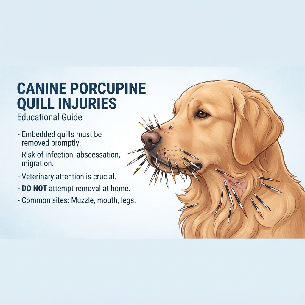
Understanding Porcupine Quill Penetration and Tissue Migration
Porcupine quill injuries are a common emergency encountered by veterinarians in regions inhabited by the North American porcupine. When a dog comes into contact with a porcupine, these specialized hairs—which are not true needles but keratinized spines—easily lodge into the dog’s skin. The fundamental danger lies in the quill's anatomy: unidirectional barbs that allow the quill to travel forward but resist backward movement. If an owner attempts to pull them out, the barbs can tear the surrounding tissue, leading to worse trauma similar to a Degloving Injury.
The Mechanics of Quill Migration
Once a quill is embedded, it does not remain stationary. The dog’s normal muscular activity causes the quill to advance through tissue. Quills are known to migrate to unexpected areas, such as the eyes, the throat, or even deep into the thoracic cavity, where they may puncture a lung or an artery. Unlike a simple Insect Sting, which causes localized reaction, quill embedment represents a dynamic, progressive internal injury.
Diagnostic Evaluation and Localization of Foreign Bodies
Veterinary diagnosis begins with a thorough physical examination to count and map the visible quills. However, because quills are often broken or buried, a simple visual inspection is never sufficient. Veterinarians must use heavy sedation or general anesthesia to properly assess the oral cavity, eyes, and deep tissues.
Advanced Imaging Techniques
When a dog is suspected of having deep-seated quills that are not visible, diagnostic imaging is required. Radiographs (X-rays) are the gold standard for detecting these foreign bodies, as they appear as radiopaque shadows. In cases where the quill has migrated near a joint or a delicate organ, a CT scan may be utilized to map the exact trajectory of the quill and prevent Joint Luxation or structural organ damage during surgery.
Surgical Extraction and Post-Operative Management
Treatment is centered on the total removal of all quills. This is not a task for home care. The process involves placing the patient under anesthesia to allow the veterinarian to thoroughly flush and explore the wounds.
Preventing Complications
After removal, the risk of a Wound Infection remains high due to the bacteria carried in by the quills. Veterinarians typically prescribe broad-spectrum antibiotics and anti-inflammatory medications. Owners must monitor for signs of abscess formation, which can occur weeks after the initial injury if a fragment was missed during the extraction process. In rare cases, if a quill has entered the chest, it may cause a Crush Injury to surrounding tissue, requiring thoracic surgery for recovery.
