

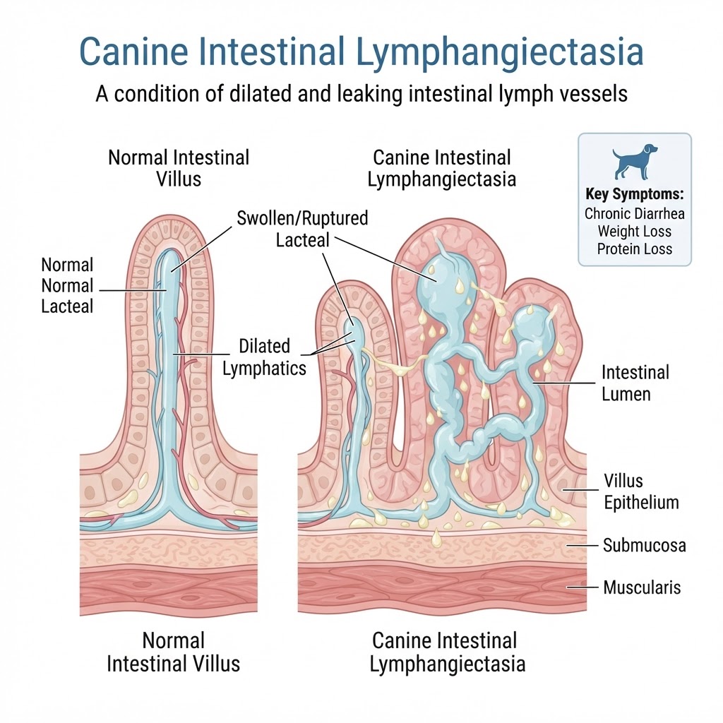
Understanding Canine Intestinal Lymphangiectasia (IL)
Intestinal lymphangiectasia is primarily a protein-losing enteropathy. In a healthy canine, the lymphatic system efficiently transports fats and immune cells from the digestive tract into the bloodstream. In cases of IL, the lacteals become permanently dilated, preventing the normal flow of lymph. This backflow results in the rupture of these fragile vessels, spilling precious proteins and lymphocytes into the intestinal tract.
Congenital vs. Acquired Lymphatic Obstruction
IL is broadly categorized based on the underlying etiology. Congenital forms occur in specific breeds and are believed to be the result of malformed lymphatic vessels present from birth. Acquired lymphangiectasia is typically a secondary consequence of chronic inflammation, infiltrative diseases, or severe gastritis that causes secondary fibrosis and lymphatic obstruction.
Recognizing Clinical Manifestations and Physiological Indicators
The symptoms of IL are often linked directly to the loss of protein and the malabsorption of nutrients. Owners should be vigilant for subtle changes in stool quality and body condition.
Primary Gastrointestinal Signs
Chronic diarrhea is the hallmark sign, though it may fluctuate in severity. Unlike a standard case of constipation, the stool in IL is often pale, voluminous, and fatty due to the inability of the intestines to properly process dietary lipids. Some dogs may also exhibit periodic bouts of vomiting or reduced appetite, which may be misdiagnosed initially as mild gastroesophageal reflux.
The Warning Signs of Hypoproteinemia (Emergency)
When serum albumin levels fall significantly, the body can no longer retain water within the blood vessels. This leads to third-spacing, where fluid leaks into the abdomen (ascites) or the chest cavity. If you observe rapid abdominal distention or increased respiratory effort, this requires immediate veterinary intervention, as it can mimic the presentation of a gastrointestinal obstruction or other life-threatening abdominal emergencies.
Diagnostic Pathways in Veterinary Medicine
Diagnosing IL requires a systematic approach to rule out other causes of protein loss. Veterinarians often begin with a complete blood count and chemistry panel to document low albumin and globulin levels. Ultrasonography is highly effective, often revealing a classic 'hyperechoic mucosal striation'—a pattern in the intestinal wall that suggests dilated lymphatics.
Definitive Diagnosis: The Histopathological Approach
While imaging provides strong clues, the definitive diagnosis is frequently achieved via endoscopic or full-thickness surgical biopsy of the intestinal wall. Pathologists look for the presence of dilated, thin-walled lymphatic vessels (lacteals) within the mucosal tissue. This process helps differentiate IL from other conditions like splenic mass or other infiltrative bowel disorders.
Comprehensive Treatment and Management Strategies
The cornerstone of treating IL is ultra-low-fat nutrition. By significantly reducing dietary fat, the pressure on the intestinal lymphatic system is minimized, which can stop the leakage of lymph fluid. Many dogs require a prescription, ultra-low-fat, hydrolyzed protein diet for the remainder of their lives.
Pharmacological Support
Management often includes immunosuppressive drugs such as prednisone or budesonide to reduce secondary inflammation. In severe cases, vitamin supplementation—specifically fat-soluble vitamins (A, D, E, K)—is necessary to prevent systemic deficiencies resulting from severe malabsorption.