

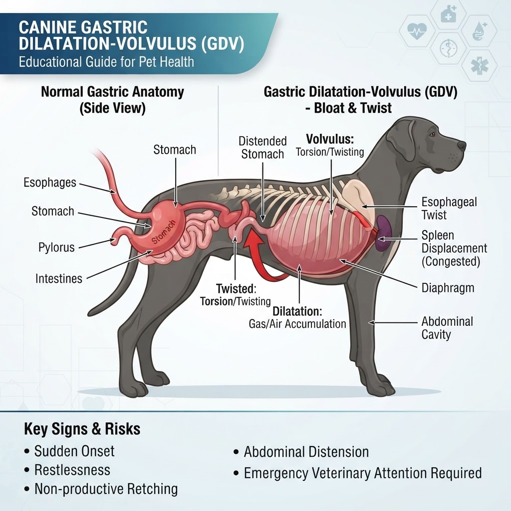
Understanding the Pathophysiology of Gastric Dilatation-Volvulus
Gastric Dilatation-Volvulus (GDV) occurs when the canine stomach undergoes massive distention followed by a rotation that traps its contents. Unlike a simple Gastrointestinal Obstruction, where the flow of contents is blocked, GDV involves a physical torsion of the pylorus and cardia, effectively sealing both ends of the organ. This prevents gas from being released via eructation or moving into the duodenum.
Mechanical Consequences and Vascular Compression
Once the stomach twists, it compresses the major blood vessels returning to the heart, specifically the caudal vena cava. This reduces the blood volume returning to the heart, which in turn drops the animal's blood pressure. Simultaneously, the stomach wall begins to lose its blood supply, leading to rapid tissue death. If the stomach was previously inflamed or affected by Enteritis, these underlying issues may exacerbate the vulnerability of the tissues.
Recognizing Clinical Indicators and Emergency Signs
Owners must recognize that GDV is an emergency that does not resolve on its own. While some dogs might show signs of Gastroesophageal Reflux, the classic signs of GDV are far more severe and persistent.
Acute Warning Signs
-
Unproductive retching: The dog attempts to vomit repeatedly but produces nothing or only small amounts of white foam.
-
Abdominal distension: A visibly tight, bloated, and hard abdomen.
-
Restlessness: Pacing, inability to get comfortable, or constant panting.
-
Hypersalivation: Drooling excessively.
-
Weakness: Collapsing or inability to stand due to cardiovascular shock.
Etiology and Risk Predispositions
While the exact triggers are debated, GDV is linked to a combination of anatomical, environmental, and dietary factors. Large and giant breeds, particularly those with deep, narrow chests, are at the highest risk. These include Great Danes, German Shepherds, and Standard Poodles.
Risk Factors and Contributing Conditions
-
Age: Older dogs are statistically more prone to developing the condition.
-
Dietary habits: Eating one large meal per day or rapid consumption of food.
-
Anatomy: A deep-chested conformation creates more 'swing' room for the stomach to twist.
-
Medical history: Dogs that have previously suffered from a Splenic Mass or Splenic Torsion are also often monitored more closely, as these conditions can alter the anatomical stability of the nearby stomach.
Veterinary Diagnostic Protocols
Veterinarians perform a rapid physical examination, often noting the 'ping' sound when percussion is applied to the abdominal wall. Radiographic imaging (X-rays) is the gold standard for confirmation, typically showing the 'double bubble' or 'Popeye arm' appearance of the stomach.
Essential Diagnostic Steps
-
Blood work: Essential to evaluate electrolyte levels, lactate (a marker of tissue damage), and overall organ function.
-
Electrocardiogram (ECG): Vital for detecting cardiac arrhythmias that frequently occur due to poor blood flow to the heart.
Surgical Intervention and Stabilization
Once diagnosed, the priority is to stabilize the patient with aggressive intravenous fluid therapy and oxygen. Surgical detorsion involves physically turning the stomach back to its normal position and assessing the tissue for necrosis. A gastropexy is performed to permanently secure the stomach to the body wall to prevent future rotation. In cases of severe damage, parts of the stomach or the entire spleen may require removal. Following surgery, the dog may require intensive care to monitor for Gastric Ulcer development or secondary complications.
