

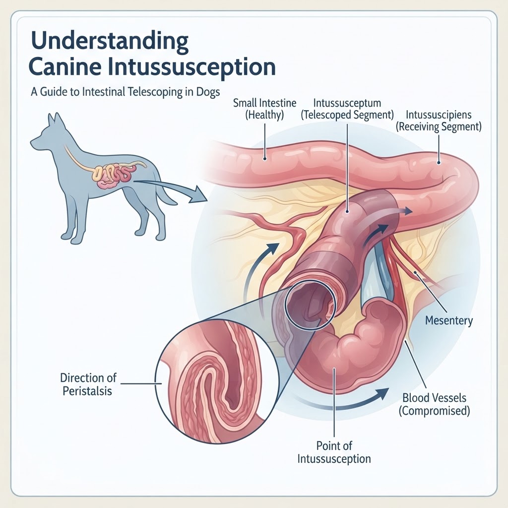
Understanding Intussusception: Bowel Telescoping
Intussusception occurs when a part of the intestine slides into an adjacent part of the intestine, much like a collapsing telescope. This movement creates an internal obstruction that blocks the passage of food and gas, while simultaneously strangulating the blood vessels supplying the affected segment.
The Anatomy of an Intussusceptum
There are several types of intussusception based on the segments involved. The most frequent location is the ileocolic junction, where the small intestine enters the large intestine. Less frequently, segments of the small intestine may telescope into themselves (jejunojejunal) or the stomach may be involved, though gastric involvement is significantly rarer. The primary mechanical risk is that the venous return from the entrapped bowel is restricted by the mesenteric tension, leading to severe ischemia. If the condition is associated with chronic enteritis, the risk of recurrence or multi-focal presentation increases.
Recognizing Clinical Indicators of Intestinal Obstruction
Dogs experiencing this condition often present with an acute onset of vomiting and lethargy. Unlike a simple gastrointestinal obstruction caused by a foreign object, intussusception involves complex vascular and inflammatory changes.
Acute Warning Signs
-
Persistent, projectile vomiting.
-
Abdominal pain that is evident when palpated.
-
Bloody, mucoid diarrhea, often described as 'raspberry jam' consistency.
-
Significant fluid loss leading to hypovolemic shock.
-
Palpable, cylindrical abdominal mass during physical exam.
Etiology: Why Does the Bowel Telescope?
In many cases, the specific trigger for an intussusception is never identified, categorized as 'idiopathic.' However, in pediatric or young dogs, intestinal parasites or severe viral enteritis often cause the hyper-motility necessary for the bowel to slide into itself. In adult dogs, the condition is more concerning because it is often secondary to an underlying mass, such as a splenic mass affecting surrounding anatomy or primary intestinal neoplasia. Additionally, chronic irritation from protein-losing enteropathy can alter intestinal motility enough to predispose an animal to this condition.
Diagnostic Pathways in Veterinary Medicine
Veterinarians must act swiftly to differentiate intussusception from other conditions like hemorrhagic gastroenteritis.
-
Physical Exam: Careful abdominal palpation can often reveal a sausage-shaped mass.
-
Radiographic Imaging: Abdominal X-rays may show a lack of gas in the colon and an unusual clustering of bowel loops.
-
Ultrasonography: The 'target' or 'donut' sign observed on ultrasound is the gold standard for confirming an intussusception.
Surgical Correction and Supportive Care
Manual reduction is the first step during surgery, where the surgeon attempts to gently 'milk' the bowel back into its normal position. If the bowel is necrotic, a resection and anastomosis are required to remove the dead tissue. Post-operative care includes intensive fluid therapy and pain management to address the systemic inflammatory response.
