

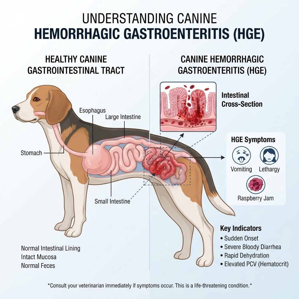
Understanding Acute Hemorrhagic Diarrhea Syndrome (AHDS)
Acute Hemorrhagic Diarrhea Syndrome (AHDS), historically known as Hemorrhagic Gastroenteritis, is a sudden, severe inflammatory condition of the canine gut. It is distinct from chronic Gastritis in its speed of onset and the severity of the bloody output. In the early stages, the barrier integrity of the intestinal mucosa is compromised, allowing for excessive leakage of fluid and protein into the digestive tract. This fluid loss results in profound hypovolemia that manifests clinically as lethargy and weak pulses.
The Pathophysiology of Vascular Fluid Loss
The hallmark of AHDS is the rapid increase in PCV, often referred to as hemoconcentration. As the blood volume decreases, the proportion of red blood cells to plasma increases, thickening the blood and making it difficult for the heart to circulate effectively. This is a primary differentiator when comparing AHDS to other causes of Gastrointestinal Obstruction, where the electrolyte imbalances are typically different in nature.
Clinical Manifestations and Emergency Indicators
Symptoms of AHDS are often described as explosive. The onset is sudden and frequently unexpected.
Primary Clinical Signs
- Profuse, watery, or bloody diarrhea resembling strawberry jam
- Repeated, forceful vomiting
- Extreme lethargy or sudden collapse
- Significant abdominal pain and guarding
- Anorexia and refusal to drink water
When to Seek Emergency Care
If you observe lethargy combined with bloody diarrhea, you must seek veterinary care immediately. AHDS can progress to death within 24 hours if the patient is not placed on intravenous fluid therapy. Do not wait for symptoms to 'run their course,' as this is not a self-limiting condition in many instances.
Diagnostic Protocols and Differential Considerations
Diagnosis is generally presumptive based on the clinical signalment and the characteristic lab findings.
Diagnostic Tools
- Complete Blood Count (CBC) with PCV/Total Solids: This is the most critical test to document the extent of hemoconcentration.
- Abdominal Ultrasound: Used to rule out a Splenic Mass or foreign body obstruction.
- Electrolyte Panel: Often reveals profound hyponatremia and hypokalemia.
Differential Diagnosis
AHDS can be easily mistaken for other severe conditions. Veterinarians must differentiate it from Parvovirus (common in younger dogs), Splenic Torsion, or severe intoxication. While Gastroesophageal Reflux often presents with vomiting, it lacks the hemorrhagic stool component of AHDS.
Therapeutic Interventions and Management
Treatment is focused on volume resuscitation. The dog is typically hospitalized for intensive intravenous fluid therapy to counteract hypovolemia.
- Intravenous Fluid Resuscitation: Crystalloids or colloids are used to expand intravascular volume.
- Antibiotic Therapy: While controversial, antibiotics like amoxicillin-clavulanate are often used to address suspected Clostridial toxins.
- Anti-emetics: Medications to control vomiting are vital to prevent further fluid loss.
Long-Term Outlook and Recovery
Once stabilized, most dogs recover within 3 to 7 days, though they may require a bland, low-fat diet for several weeks to allow the intestinal mucosa to fully repair.
