

Understanding Acquired Portosystemic Shunts in Dogs
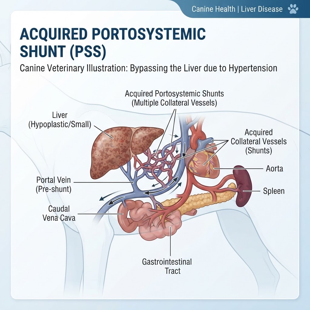
A portosystemic shunt (PSS) is an abnormal blood vessel that allows blood from the gastrointestinal tract and spleen to bypass the liver and flow directly into the systemic circulation. While congenital PSS are present from birth due to developmental errors, acquired PSS develop later in life. They are typically a consequence of increased pressure within the portal vein, often secondary to underlying liver disease, cirrhosis, or certain cardiac conditions that impair venous return to the heart. The liver normally filters toxins and metabolizes nutrients from blood returning from the digestive system; when blood bypasses this crucial detoxification and metabolic processing, toxins accumulate in the bloodstream, affecting various organ systems, most notably the brain.
Recognizing Clinical Signs of Portosystemic Shunt
The clinical presentation of acquired portosystemic shunts in dogs is highly variable, depending on the severity of the shunt, the degree of underlying liver compromise, and the accumulation of toxins. Owners may notice a gradual onset of nonspecific signs or sudden, severe episodes. Common indicators include:
- Neurological Abnormalities (Hepatic Encephalopathy): This is a hallmark of PSS and results from the accumulation of toxins, especially ammonia, in the brain. Signs can range from mild disorientation, lethargy, and unsteadiness to more severe symptoms like head pressing, seizures, tremors, circling, apparent blindness, stupor, or coma.
- Gastrointestinal Upset: Vomiting, diarrhea, poor appetite, and failure to gain weight or maintain body condition are frequently observed.
- Urinary Tract Issues: Increased thirst (polydipsia) and increased urination (polyuria) can occur. In some cases, ammonium urate uroliths (bladder stones) may form due to altered urine composition.
- Poor Growth and Development: Young dogs with PSS may exhibit stunted growth.
- Behavioral Changes: Disorientation, depression, or aggression can be signs of hepatic encephalopathy.
- Weakness and Lethargy: General malaise and reduced activity levels.
- Poor Coat Quality: A dull, unkempt coat may indicate nutritional deficiencies or systemic illness.
- Abdominal Distension: Fluid accumulation in the abdomen (ascites) can occur in advanced cases, often due to compromised liver function and low protein levels.
Warning Signs of Hepatic Encephalopathy Crisis (Emergency)
Sudden or severe neurological deterioration, including unresponsiveness, repeated seizures, or profound weakness, constitutes a medical emergency requiring immediate veterinary attention.
Etiology and Risk Factors for Acquired Shunting
Unlike congenital PSS, which stem from incomplete closure of fetal vessels, acquired shunts are a pathological adaptation. The primary driver is usually increased resistance to blood flow within the liver or the portal venous system. This can be caused by:
- Chronic Liver Disease: Conditions such as chronic hepatitis, fibrosis, and cirrhosis lead to scarring and narrowing of the liver's blood vessels, impeding portal blood flow.
- Cirrhosis: Advanced scarring of the liver tissue significantly increases resistance.
- Vascular Obstruction: Tumors or thrombi within the portal vein or hepatic vessels can obstruct flow.
- Cardiac Disease: Conditions leading to increased pressure in the vena cava, such as right-sided heart failure or constrictive pericarditis, can impede blood outflow from the liver.
While specific breed predispositions are more pronounced for congenital PSS, any dog developing conditions that cause significant portal hypertension or liver damage is at risk for acquired shunts.
Diagnostic Pathways for Portosystemic Shunt
Diagnosing acquired portosystemic shunts involves a multi-faceted approach by veterinarians to confirm the shunt's presence, assess its severity, and identify any underlying causes.
Initial Physical Examination Findings
A thorough physical exam may reveal findings such as a palpable abdominal fluid wave (ascites), poor body condition, signs of neurological impairment, or a small-sized liver (sometimes). The veterinarian will assess vital signs, listen to the heart and lungs, and palpate the abdomen.
Laboratory Investigations for Hepatic Function
Blood work is crucial. Standard tests include:
- Complete Blood Count (CBC): May show anemia or signs of infection.
- Serum Biochemistry Panel: Crucial for assessing liver enzymes (ALT, AST, ALP, GGT), bile acids (pre
- and post-prandial), albumin, globulins, blood urea nitrogen (BUN), and electrolytes. Elevated pre-prandial and particularly post-prandial bile acids are highly suggestive of a shunt or significant liver dysfunction. Low albumin and urea can indicate poor liver synthetic function.
- Urinalysis: To evaluate urine concentration and check for crystals or stones, particularly ammonium urate stones.
- Ammonia Levels: Elevated blood ammonia levels, especially when correlated with clinical signs, strongly support hepatic encephalopathy.
Advanced Imaging Modalities
- Abdominal Ultrasound with Doppler: This is often the primary imaging modality. It allows visualization of the liver size and texture, identification of any intra-abdominal masses, and importantly, Doppler assessment of blood flow. It can often detect abnormal vascular connections entering the vena cava or azygous vein, bypassing the liver.
- Contrast-Enhanced CT Angiography or MR Angiography: These advanced imaging techniques provide detailed, three-dimensional visualization of the vasculature and are considered the gold standard for definitively identifying and characterizing portosystemic shunts, including their exact location, size, and any associated abnormalities.
- Plain Radiographs (X-rays): May show a small liver, abnormal vasculature (sometimes), or uroliths, but are less definitive for shunt identification.
Diagnostic Imaging for Vascular Anomalies
Specialized imaging like CT angiography or MR angiography is critical for precise anatomical definition of the shunt tract and can guide surgical planning.
Management and Treatment Strategies
Treatment for acquired portosystemic shunts focuses on managing the clinical signs, addressing the underlying cause, and in some cases, attempting to reduce the shunt's effect.
Medical Management of Clinical Signs
This is often the cornerstone of treatment, especially when surgical intervention is not feasible or the underlying cause cannot be fully corrected.
- Dietary Modification: A specialized veterinary diet low in protein but of high biological value (e.g., using dairy or soy protein) is often recommended to reduce ammonia production in the gut. Lactulose, a non-absorbable sugar, is commonly prescribed to bind ammonia in the colon and promote its excretion, thus reducing systemic absorption. Probiotics may also be beneficial for gut health.
- Antibiotics: Certain antibiotics like metronidazole or neomycin can reduce ammonia-producing bacteria in the gut, further aiding in the management of hepatic encephalopathy.
- Fluid Therapy: Intravenous fluids may be necessary to correct dehydration and electrolyte imbalances, especially during episodes of vomiting or diarrhea.
- Medications for Seizures: Anticonvulsant medications (e.g., phenobarbital, levetiracetam) are used to control seizures associated with hepatic encephalopathy.
- Diuretics and Management of Ascites: If ascites develops, diuretics may be used to help remove excess fluid, alongside measures to increase serum albumin if needed.
Surgical Intervention Considerations
Surgical correction of acquired portosystemic shunts is often more complex and less frequently attempted than for congenital shunts. This is because acquired shunts are usually more numerous, tortuous, and diffuse, often developing as a widespread compensatory response to severe, chronic liver disease. If a single, discrete acquired shunt is identified and the underlying liver condition is not too advanced, surgical ligation or attenuation might be considered. However, more commonly, the focus remains on medical management. Procedures may involve surgically placing a band or stent to gradually reduce blood flow, or more complex interventional radiology techniques.
Liver Support and Supportive Care
Supportive care involves managing symptoms, ensuring adequate nutrition, and preventing complications. This includes monitoring for signs of hepatic failure and providing symptomatic relief for issues like nausea or pain.
Post-Treatment Recovery and Long-Term Outlook
The recovery and long-term prognosis for dogs with acquired portosystemic shunts are highly dependent on the underlying cause, the severity of the liver disease, and the success of management strategies.
Recovery Phases
-
Phase 1: Stabilization and Symptom Control: This initial phase focuses on managing acute clinical signs, particularly neurological episodes or gastrointestinal upset, through intensive medical therapy. Bloodwork is re-evaluated to monitor organ function and response to treatment.
-
*Timeframe:
-
Days to weeks.
-
Phase 2: Medical Management Optimization: Once stabilized, the focus shifts to long-term medical management. This involves fine-tuning diet, medications (lactulose, antibiotics), and monitoring for any recurrence or worsening of signs.
-
*Timeframe:
-
Weeks to months.
-
Phase 3: Long-Term Monitoring and Lifestyle Adjustments: This is an ongoing phase where the dog requires lifelong management. Regular veterinary check-ups, including bloodwork and potentially imaging, are essential to monitor liver health and shunt status. Owners must adhere strictly to dietary recommendations and medication regimens.
-
*Timeframe:
-
Lifelong.
Lifelong Management of Shunt Effects
Dogs with acquired PSS will likely require ongoing medical and dietary management for the remainder of their lives to control symptoms and support liver function.
Prevention Strategies for Acquired Shunts
Direct prevention of acquired portosystemic shunts is difficult because they are usually a consequence of other diseases. The focus is on preventing or aggressively managing the conditions that lead to their development:
- Preventing Liver Disease: Maintaining a healthy weight, providing a balanced diet, avoiding toxins (certain plants, chemicals, medications), and prompt treatment of infections that can affect the liver can help protect liver health.
- Early Detection and Management of Underlying Conditions: If a dog develops conditions known to cause portal hypertension or severe liver damage (e.g., chronic hepatitis, heart failure), prompt and aggressive veterinary care is crucial. Early intervention may slow disease progression and potentially reduce the risk or severity of shunt formation.
When to Seek Emergency Veterinary Care
Immediate emergency veterinary care is required if a dog exhibits any of the following signs, which can indicate a severe complication or crisis related to a portosystemic shunt:
- Severe Neurological Signs: Repeated seizures, coma, profound weakness, inability to stand, or sudden disorientation.
- Gastrointestinal Crisis: Persistent vomiting that prevents fluid retention, severe abdominal pain.
- Respiratory Distress: Difficulty breathing, which could be related to ascites or other systemic complications.
- Collapse or Unconsciousness: Any sudden loss of consciousness.
For less severe or persistent symptoms like mild lethargy, decreased appetite, or occasional vomiting, a prompt appointment with your veterinarian is recommended for evaluation and diagnosis.
Differential Diagnoses for Shunt-Like Symptoms
When evaluating a dog with signs suggestive of a portosystemic shunt, veterinarians consider other conditions that can mimic these symptoms:
- Congenital Portosystemic Shunt: Especially in younger dogs, distinguishing between congenital and acquired shunts is important.
- Hepatic Failure (acquired): Other causes of severe liver dysfunction unrelated to shunts, such as toxicity, infectious hepatitis, or bile duct obstruction.
- Gastrointestinal Obstruction: Vomiting, anorexia, and abdominal pain can mimic shunt signs, particularly if enteritis is present.
- Nephrotic Syndrome: Can cause hypoalbuminemia leading to edema and ascites.
- Congestive Heart Failure: Can lead to ascites and general weakness.
- Certain Toxicoses: Ingesting toxins can cause neurological signs and gastrointestinal upset.
- Metabolic Disorders: Such as Addison's disease or severe electrolyte imbalances.
Breed Predispositions for Shunts
While acquired shunts are less tied to specific breeds than congenital ones, certain breeds are predisposed to conditions that can lead to acquired shunts. For instance, breeds prone to chronic hepatitis (e.g., Bedlington Terriers, Doberman Pinschers, Labrador Retrievers) are at higher risk. Similarly, breeds susceptible to heart disease may be at increased risk for shunts secondary to cardiac compromise. Ultimately, any dog can develop acquired shunts if the underlying conditions are present.
