

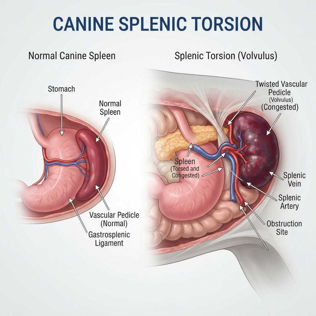
Understanding Splenic Torsion: Mechanisms of Vascular Occlusion
Splenic torsion is a condition characterized by the rotation of the spleen and its primary blood vessels. This rotation results in a mechanical strangulation of the splenic vein and artery. Without effective circulation, the spleen becomes massively engorged with blood, leading to tissue death and the release of inflammatory mediators. This condition often resembles Gastrointestinal Obstruction in terms of its acute abdominal presentation.
The Role of the Splenic Pedicle
The spleen is held in place by several ligaments; however, in some individuals, these ligaments are lax, allowing for excessive mobility. When the spleen rotates, the pedicle twists, effectively creating a vascular knot. This prevents blood from leaving the organ (venous congestion) while simultaneously blocking oxygenated blood from entering, leading to ischemia.
Recognizing Clinical Signs and Emergency Indicators
Clinical signs of splenic torsion are often nonspecific but intense. Owners may notice sudden abdominal discomfort, restlessness, and non-productive retching. Unlike Gastritis, which often presents with intermittent vomiting, splenic torsion involves profound lethargy and progressive weakness.
Acute Abdominal Pain and Distension
-
Extreme restlessness and inability to find a comfortable position.
-
Visible distension of the cranial abdomen.
-
Pale mucous membranes signaling the onset of shock.
-
Rapid, labored breathing as the animal compensates for severe pain.
Etiology and Breed Predisposition
While the exact cause of idiopathic torsion remains unknown, it is frequently associated with anatomic abnormalities of the ligaments supporting the spleen. It is heavily linked to deep-chested breeds such as Great Danes, German Shepherds, and Standard Poodles.
Environmental and Secondary Risk Factors
-
Concurrent gastrointestinal distress or chronic Enteritis.
-
Increased abdominal pressure due to chronic Constipation.
-
History of gastric dilation, which shifts surrounding anatomy and provides the mechanical space for rotation.
Diagnostic Protocols
Veterinarians diagnose this condition using a combination of physical examination, diagnostic imaging, and baseline blood work. The physical exam often reveals a large, firm mass in the abdomen. Ultrasound imaging is the gold standard, as it can visualize the 'whirl sign'—a specific pattern created by the twisted vessels in the splenic pedicle.
Surgical Intervention and Supportive Care
Treatment is exclusively surgical. A splenectomy, the surgical removal of the spleen, is required to prevent further systemic poisoning from the necrotic tissue. Supportive care during surgery includes aggressive fluid therapy to treat hypotension and, if necessary, the administration of blood products.
Post-Surgical Considerations
Following a splenectomy, patients must be monitored for arrhythmias, which are common complications during the recovery phase. Careful observation for signs of Peritonitis is crucial as the animal heals. Prognosis is generally good if surgery is performed before the onset of profound shock or septicemia.