

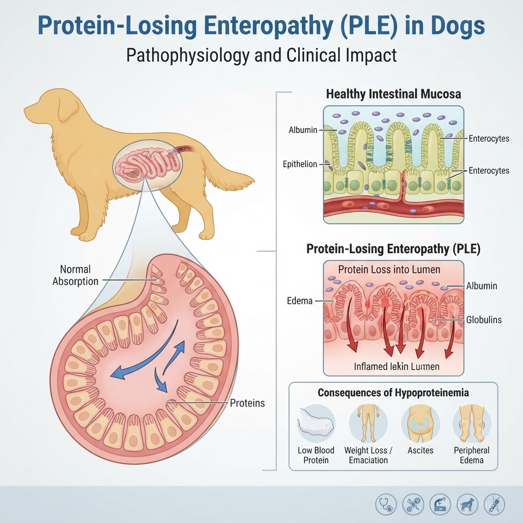
Pathophysiology of Protein-Losing Enteropathy
Protein-Losing Enteropathy (PLE) is not a single disease entity, but rather a functional consequence of various gastrointestinal disorders that result in protein loss. When the gastrointestinal barrier is breached through enteritis or lymphocytic-plasmacytic infiltration, protein molecules leak into the gut lumen. This is often exacerbated by lymphangiectasia, where obstructed lymphatic vessels rupture, dumping protein-rich lymph into the intestines.
Impact on Oncotic Pressure and Fluid Balance
As serum albumin levels drop, the oncotic pressure of the blood declines significantly. This creates an imbalance where fluid leaves the intravascular space and accumulates in the abdomen (ascites) or legs (pitting edema). Unlike gastritis, which involves acute inflammation of the stomach lining, PLE is typically a chronic, systemic wasting process that affects the entire intestinal absorptive surface.
Clinical Manifestations and Diagnostic Indicators
Recognizing the subtle signs of PLE early is critical for owner intervention. While many dogs initially present with mild weight loss, the condition can quickly progress to systemic weakness.
Common Symptom Clusters
-
Persistent, watery diarrhea
-
Severe weight loss despite a normal or ravenous appetite
-
Fluid retention manifested as bloated abdomen
-
Lethargy and reduced exercise tolerance
-
Poor coat quality due to malnutrition
Warning Signs of Critical Deterioration
If a dog with diagnosed PLE begins to show extreme respiratory distress (dyspnea), this may indicate pleural effusion requiring emergency drainage. Owners should be wary of any sudden changes in gum color or signs of gastrointestinal obstruction, which can occasionally mimic or complicate PLE cases.
Diagnostic Framework for Enteric Protein Loss
Veterinarians utilize a tiered approach to reach a definitive diagnosis. Initially, complete blood counts (CBC) and serum chemistry panels often reveal panhypoproteinemia (low albumin and low globulin). Unlike other diseases where only albumin is low, the combined decrease is a hallmark of protein-losing conditions.
Imaging and Biopsy Procedures
-
Abdominal Ultrasound: Crucial for identifying thickening of the intestinal walls or enlarged mesenteric lymph nodes.
-
Endoscopic Biopsy: This is the gold standard for diagnosing mucosal disease, allowing for the visual assessment of the gut lining.
-
Serum Cobalamin/Folate Levels: Often low in PLE patients due to malabsorption.
Therapeutic Approaches and Management Protocols
Treatment is heavily dependent on the biopsy findings. If IBD is the culprit, high-dose corticosteroids or secondary immunosuppressants are often utilized. If lymphangiectasia is suspected, a ultra-low-fat diet is the cornerstone of therapy to reduce lymphatic pressure in the intestinal walls.
Nutritional Support and Monitoring
Dietary management is life-long. Many patients thrive on hydrolyzed protein diets or home-cooked low-fat formulations. If left untreated, the risk of developing secondary complications like portosystemic shunt (due to portal hypertension) increases, though this is rare compared to the primary effects of hypoalbuminemia.
